Understanding cellular ultrastructure in 3D context with ZEISS Volutome
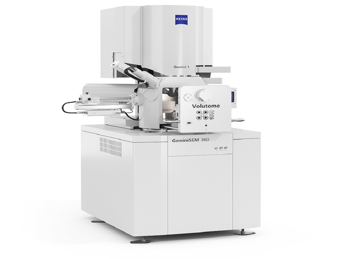
An in-chamber ultramicrotome for ZEISS field emission scanning electron microscopes (FE-SEM), the ZEISS Volutome is a recently released product. It is intended for use in the life sciences to create 3D images of the ultrastructure of biological material implanted in resin.
With several techniques collectively referred to as volume EM (vEM), scanning electron microscopy (SEM) in general can be utilized to investigate complex, ultrastructural 3D information. For researchers who seek simple sample preparation combined with a fully automated imaging technique that enables the gathering of huge volume datasets, serial block-face SEM (SBF-SEM) is the preferred vEM technology.
The ZEISS Volutome is a hardware-to-software end-to-end serial block-face imaging solution with image processing, segmentation, and visualization capabilities. The 3D FE-SEM may be converted into a regular FE-SEM by simply swapping out the ultramicrotome, making the system adaptable to a variety of environments.
Saving time with automated cutting, image acquisition, and pre-processing
Imaging biological structures in vast volumes at high resolution might take days. As a result, steady acquisition conditions for a long time are necessary for SBF-SEM. Highly automated, unsupervised cutting and imaging are possible with the ZEISS Volutome. Images are simultaneously pre-calculated for stitching and z-stack alignment during image acquisition. With just one click, the user can access the results.
3D imaging in every way
The new high-speed, high-sensitivity detector for serial block-face imaging is included with the ZEISS Volutome: High contrast images are possible even at low acceleration voltages because of ZEISS Volume BSD. Charge-prone samples can be conveniently and without compromise photographed using the ZEISS proprietary Focal Charge Compensation in conjunction with charge neutralization at the block face. Additionally, the integrated ZEISS Volutome stage makes it easier to acquire a huge volume of EM information.
Applications
ZEISS serial block-face imaging Any biological material that has been processed for electron microscopy can be studied using the volume to examine cellular structures. Several well-known study areas include plant science, cell biology, and imaging of tissues in general.
Researchers across many neuroscience disciplines continue to use serial block-face SEM to gain a better knowledge of neural connections and signaling pathways, whether they are working on basic questions in developmental, cellular, or molecular neuroscience or doing practical studies on aging or neurodegenerative illnesses.
To view the ultrastructure of cells and cellular components, examine morphology, and quantify structures of interest in cell biology, high-resolution imaging is required.
Multiple blocks face SEM uncovers ultrastructural alterations in plants brought on by genetic mutations, drought, pollution, or climate change. These variables affect how well-off or ill plants are, which in turn affects crop yield, food production, and eventually, human health.
Serial block-face imaging enables huge sample tissue volumes to be photographed and analyzed within a broader 3D environment, regardless of whether researchers work with tumors and biopsies, organ or tissue slices, organoids, embryos of model organisms, and more.
One contact for serial block-face imaging
ZEISS provides an end-to-end solution from hardware to software and has years of experience in vEM involving serial block-facing imaging. Based on the foundational research technology of the Max-Planck Institute for Neurobiology in Martinsried, Germany, ZEISS Volutome was developed. To deliver solutions that are suited to the demands of the users, ZEISS maintains regular interaction with the research community.
Click on the following link Metrologically Speaking to read more such news about the Metrology Industry.


